Bigotry: The Dark Danger
The Miracle in the Cell Membrane

DOWNLOAD THE BOOK
CHAPTERS OF THE BOOK
- Foreword
- Introduction: The Consciousness Beyond Matter and The Col Lapse of Mechanism
- The Miniature Factories Comprising Our Bodies: The Cells
- The Cell's Complex Structure Can not Be Explained in Terms of Coincidence
- Superior Creation in The Structure of The Cell Membrane
- Complex Transportation Systems in The Cell Membrane
- The Selective Permeability of The Protein Channels in The Cell Membrane
- Selectivity in Nerve Cells
- Signal Selection in Traffic of Data Among Cells
- Selection in The Immune System's Cells
- Vital Selections in The Blood
- The Importance of The Creation in The Cell Membrane in Terms of Multi-Cellularity
- The Delicate Balance in Substances Selected in The Body
- The Cell Membrane Invalidates Claims of The Theory of Evolution
- Conclusion: Allah Pervades Everywhere With His Wisdom
< <
8 / total: 15
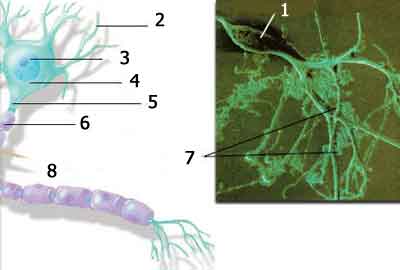
Selectivity in Nerve CellsIn contrast to other cells, a nerve cell or neuron possesses sections known as dendrites and axons. A dendrite consists of a large number of short protrusions like roots or branches and plays a role in receiving stimuli from other neurons and cells and transmitting them to the cell body. Axons are long, thin fibers emerging from the body of the cell, consisting of one single part along which stimuli are sent, serving to transmit messages to the brain. Nerve cells thus form a dense network of long chains.
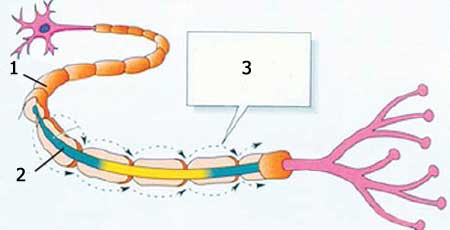
Every cell has an electrical charge around its membrane. Every neuron resembles a miniatured biological battery ready to discharge its energy. The ions, electrically charged molecules both inside and outside every nerve cell, set up a difference of electrical charges along the length of the cell membrane. To send a stimulus, human neurons require an average electrical charge of minus 50 millivolts (One millivolt is 1 thousandth of a volt).50 At this point, the nerve signal is transmitted by the axon. After every nerve signal passes, potassium ions flow from the cell membrane. After every signal, the neuron must be recharged. In order to do that, it takes ions back until the potential level is regained.
For a neuron to send a signal takes around 1 thousandth of a second, so in one second, it is possible to send at most 1,000 signals. In general, however, only 300 to 400 signals take place every second. Human nerve cells are of different lengths.51 The transmission between cells takes place at speeds between 3 and 100 meters (9.8 and 328 feet) per second. 52 Professor Peter Suckling, a neurophysicist at the Downstate Medical Center, speaks of the cell membrane with great amazement: "This very thin membrane can sustain an electrical tension better than most insulators. The insulation strength is high. It has to be strong; it's so very thin." 53 Nerve cells are able to communicate as a result of the electricity produced in the cell membrane, to transmit information from one place to another, and ensure the healthy operation of the body's functions. In addition, these electrical messages produced in the cell reach their intended destinations, carrying a message for the recipient cell. Every cell knows the meaning of the message that reaches it, and begins operating accordingly. Were there no such flawlessly operating systems among the cells, it would be impossible for any living thing to maintain vital functions. That being so, then how did such a flawless system requiring consciousness and intelligence arise? It is of course impossible to maintain that unconscious masses of atoms and molecules took a decision to form cells and that such a system later spontaneously came into being among those cells. The existence of such a conscious system proves that living things were created. The magnificent creation in such microscopic dimensions that astonishes scientists belongs to our Lord, the Creator of all. "Is He Who creates like him who does not create? So will you not pay heed?" (Surat an-Nahl, 17)
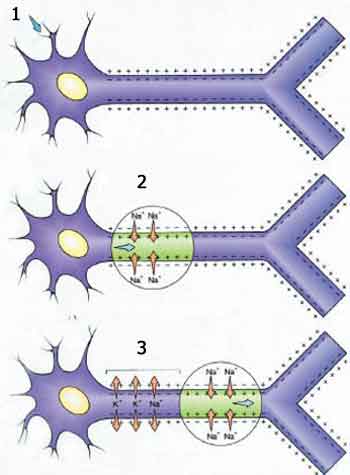
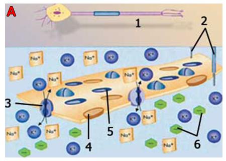
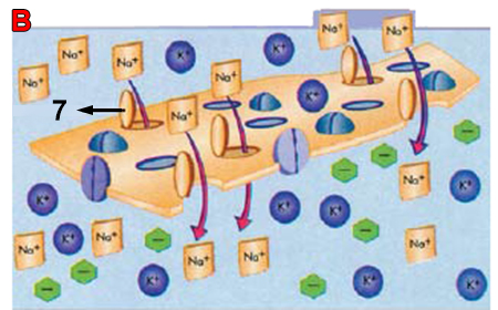
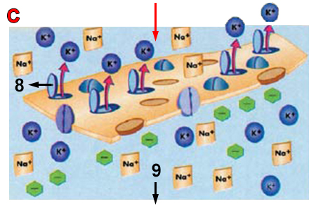
The Neuron at RestWhen a nerve cell is not transmitting a signal, it is at rest, but is not entirely immobile. It must be ready to forward messages that may arrive at any moment from neighboring cells. A neuron at rest must always be polarized, so that the fluid within it is more negatively charged than the fluid outside. Along its membrane, a nerve cell has an electrical potential of approximately 70 millivolts. This is called the membrane potential or resting potential. Though this appears very small, it means that a tiny cell produces up to 1/20th of the voltage of a flashlight battery, and the axon holds the potential for electrical activity along the membrane. How does this resting potential come about? How is it conserved? Outside the axon, there are sodium (Na+) and chloride (Cl-) ions. Inside are charged proteins and potassium (K+) ions. The electrical imbalance between the cell membrane and the exterior forms a resting potential along the membrane, an imbalance formed by the charged ions that lets the cell membrane be selectively permeable for different ions. Sodium, potassium and chloride ions pass through the cell membrane, but the passage of highly-charged proteins is restricted—and thus, so is the formation of electrical potential. However, selective permeability cannot be the sole answer, because the number of potassium (K+) ions in the cell is always greater than the number of sodium (Na+) ions. Furthermore there are more sodium (Na+) ions outside the cell than there are potassium (K+) ions. For the requisite ion balance to be established, the densities inside the nerve cell must be reversed. The cell achieves this by using the kind of ion pump we touched on earlier. The sodium-potassium pump, a protein molecule, forms a channel in the cell membrane, taking its energy from ATP (or adenosine triphosphate, the cellular energy molecule used directly by living things) and takes in potassium (K+) ions as it expels sodium (Na+) ions. In this way, the correct ion proportions are maintained inside and outside the cell. There are between 100 and 200 sodium-potassium pumps on every square micrometer on the cell membrane surface. Each one expels 200 sodium ions a second, and admits 130 potassium ions. Movement Potential and Signal TransportationThe signal begins when a neuron is stimulated by another neuron or its environment. Immediately afterward, the signal acts along the length of the axon, causing the cell membrane's potential to suddenly reverse. In the cell membrane, there are thousands of protein channels or gates for the passage of ions, but these gates are generally closed. In the event of a signal, the sodium channels open, and positively charged sodium ions flow in. Temporarily, therefore, the interior of the cell membrane has a greater positive charge than the outside, and the resting potential is reversed, raising the cell membrane's potential to +50 millivolts. The reversal of these charges is called movement potential. During movement potential, the potassium gates open, and positively charged potassium ions flow out. This again balances the resting potential, so that the neuron's interior is again negatively charged and the exterior positively charged. One single nerve impulse triggers this entire process. You can therefore compare the signals to dominos. As one domino falls over, it pushes the one next to it. But in this case, as the signal passes, the "dominos" right themselves again and stand up, preparing themselves for a subsequent movement potential.
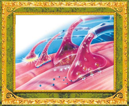
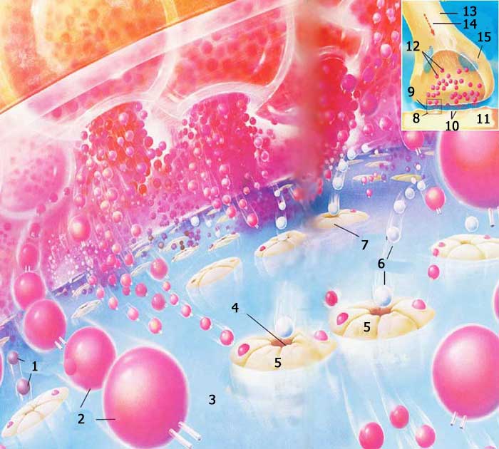
Synapse PathsThe human nervous system is a complex network consisting of billions of nerve cells that establish communication among themselves and other cells in the body by means of synapses—small parts of the nerve cells that approach one another very closely but never actually touch. Since they never come into contact with one another, signals do not pass from one cell to another directly, but are carried through the gaps by chemical neurotransmitters. When a signal reaches the transmitting cell, that cell causes some neurotransmitters to be secreted into the extracellular space. At this the neurotransmitter molecules diffuse in this space—passing directly into a less dense environment—and attach to the receptor protein molecules on the second cell. Since there are many kinds of neurotransmitter and receptor molecules, the synapse can cross very quickly (in 1/1000th of a second) or rather slowly (1/100th of a second). Chemical agents set the second cell in action, or else halt it. Therefore, synapses serve to alter the information in the nervous system or set it in motion. Because of these properties, the function of synapses in the brain is connected with learning and memory. As neurons transmit messages by means of the synapses, they exchange chemical signals. The nerve cells in your brain have 100 trillion connections, where there is a constant and enormous molecular traffic. The electrically-charged chemicals known as ions and large and small varieties of proteins tell this traffic when to flow, and when to halt.
3. Brain Cell Selectivity: "The Blood-Brain Barrier"
In the brain, special sentries admit the necessary nutrients in the blood but keep out substances that would keep nerve cells from functioning. These sentries form a barrier between the brain's nerve tissues and prevent substances in the blood from entering. This barrier is constituted by endothelium cells that line the brain's blood vessels. The barrier's importance stems from the fact that nerve cells need a specific chemical environment. Lacking such an obstacle, when you ate foods that increase the concentrations of glucose, amino acids, hormones or other compounds, or when you engaged in exercise, nerve functions would move out of control and would even suffer seizures. In the brain, countless capillaries bring nutrients and carry away waste products. The brain's endothelium cells have special connections that prevent substances in the blood from passing through the cell membrane and reaching the nerve tissue. For that reason, the endothelium cells are almost totally impermeable. But did the barrier not allow anything pass, then the brain would be deprived of the needed oxygen, glucose and amino acids and would die. Yet the blood-brain barrier's special mechanisms keep unwanted substances out, but carry vital molecules to the brain. Molecules soluble in fat can generally pass through the blood-brain barrier immediately. These include nicotine, ethanol and heroin. However, if charged molecules are not soluble in fat and must depend on special transport systems, they enter the brain very slowly, or not at all. These may be large molecules such as proteins or small ones like sodium. Glucose—the brain's main source of the energy it needs—and the amino acids that it cannot produce itself, are not soluble in fat. Therefore, these substances are carried through the cell membrane by particular transporters. The human brain uses more than 120 grams of glucose a day. But since it cannot store more than 2 grams, glucose must be constantly pass through the barrier. In the light of this need, a great many transporters in every endothelium cell allow it to absorb large quantities of glucose from the blood. The glucose transport system is the body's hardest-working transport system. The cell itself uses only a very small part of this sugar, and the remainder is transferred to the brain. Yet the structure of the transport molecules is still a mystery to scientists. In all likelihood, the transporters are one or more proteins that open the channels so as to permit glucose to pass through the cell membrane. Amino acid transport systems are far more complex, because every one of the 20 amino acids has a different molecular structure. These can be grouped into four classes depending on their chemical properties: large neutral, small neutral, basic and acidic. Each category has its own transport system. As with glucose transporters, large neutral amino acid transporters lie on both sides of the barrier, and amino acids can thus enter and leave the brain. Small neutral amino acids can be synthesized by the brain cells, so there is no need for them to be transported to the brain. The idea of the blood-brain barrier was first advanced by the German bacteriologist Paul Ehrlich at the end of the 19th century. It was possible to be proved, however, only with the development of the electron microscope in the 1950s. Although in appearance capillaries in the brain resemble the veins in other parts of the body, they possess different features. First of all, the connections between cells in the capillary vein in the brain are exceedingly dense. At their connection points, cell membranes are attached to one another just like a zipper. Unions between the endothelium cells in the capillary vessels in the other parts of the body have spaces. Second, in cells in the brain's capillary vessels, there are very few pinocytosis sacs that help transport fluids and solutes through the cell membrane. In cells outside the brain, however, these sacs are widespread.
The importance of the barrierWe can better understand the importance of the blood-brain barrier from diseases that occur in that barrier's absence. Tumors, brain-tissue defects and edema—swellings due to the accumulation of fluids and proteins—cause this barrier to collapse. Since fewer sacs form in the walls of the endothelium cells, leakage begins, or the tight bonds between the cells are loosened. Damage to the barrier leads to the accumulation of fluid and lead poisoning in brain tissue. The metal first enters the endothelium cells and then the astrocytes. After the lead has damaged the barrier, the brain is more vulnerable to other substances. The blood-brain barrier is no longer considered a passive structure, but a dynamic interface between the blood and the brain. Yet our understanding of its maintenance and transport mechanisms is still incomplete. 54 Every portion of our bodies has been specially created for life. These parts, only a few of which we shall look at in this book, have occupied scientists for decades and contain mechanisms that astound investigators. Why is this blood-brain barrier in exactly the right place only and not among cells in any other part of the body? How do the cells know that the brain needs a stable environment, and so entry to and exit from its cells must be more tightly controlled? No doubt the cells themselves do not decide to form a barrier and then construct one in the membranes. It is out of the question for this to be the work of coincidence. The barrier in the brain serves a vital purpose, and this complex structure is directed towards that end. Therefore, Darwinists hoping to account for the origin of life in terms of coincidental mechanisms are once again at a dead end. Even if all the body's complex systems existed but only this barrier in the brain cells did not, then you could not survive. That being so this same barrier must have been present, along with all its systems, from the very outset. Gradual development, the basis of the Darwinists' claims, is again invalidated, as this example shows. As an indicator of planned structure, this precautionary measure taken for human beings is one of the countless proofs of the existence of Allah. "Then inquire of them: Is it they who are stronger in structure or other things We have created? We created them from sticky clay. No wonder you are surprised as they laugh with scorn!" (Surat as-Saffat, 11-12)
Footnotes49. http://www.abe.msstate.edu/classes/abe4323/2002/cells/cells_ques.html 50. http://www.noteaccess.com/APPROACHES/ArtEd/ChildDev/ 1cNeurons.htm; [Coon, Dennis. Introduction to Psychology, Exploration and Application. St. Paul: West Publishing Company, 1989.] 51. Ibid. 52. http://www.remarkablemedicine. com/Medicine/bodyelectricity.html 53. Ibid. 54. N. Ramlakhan, J. Altman, "Breaching the blood-brain barrier," New Scientist, No.128, 1744, 24 November, 1990.
|
|||||||||||||||||||||||||||||||||||||||||||||||||||||||||||


8 / total 15
You can read Harun Yahya's book The Miracle in the Cell Membrane online, share it on social networks such as Facebook and Twitter, download it to your computer, use it in your homework and theses, and publish, copy or reproduce it on your own web sites or blogs without paying any copyright fee, so long as you acknowledge this site as the reference.